

What Vitamin Deficiency Effects Nails
Department of Dermatology, Venereology, and Leprology, Postgraduate Institute of Medical Education and Research, Chandigarh, India
Correspondence Address:
Dipankar De
Department of Dermatology, Venereology, and Leprology, Postgraduate Found of Medical Education and Enquiry, Chandigarh
India
Nails are not only an important attribute of the external appearance, they are also mirrors of the internal constitution and nutritional condition. Nail changes in nutritional deficiencies are generally minor and non-specific. It is often difficult to suspect a nutritional deficiency state just by observing boom changes. In this article, nosotros accept discussed the bones contents of the nail plate and the different nail changes that can exist observed in various nutritional deficiency states.
The nail appliance consists of a horny "dead" product, the blast plate, and four specialized epithelia: the proximal nail fold, the nail matrix, the nail bed, and the hyponychium. [1] The bulk of the nail plate is constituted by hair type (difficult) keratins which contain 80% to 90% of the nail plate. Epithelial blazon keratins account for 10% to 20%. The overall sulphur content is approximately 10% past weight. The disulfide bonds of cystine in the matrix proteins contribute maximally to smash hardness past gluing the keratin fibers together. Calcium does not contribute to nail hardness and makes upwards only 0.ii% of the boom plate past weight. The lipid content is relatively low compared with the lipid content of the stratum corneum. Glycolic and stearic acids are boom plate lipids and their presence contributes to the h2o resistance. Despite the water resistance, the hydration status of the nail plate is another cistron determining its hardness. The smash plate′s h2o content tin vary profoundly, with normal content being eighteen%. Nails become brittle when the water content is less than 16% and become soft when greater than 25%. Minerals are another important attribute of the nail plate′s composition; mainly, magnesium, calcium, iron, zinc, sodium, and copper. [2]
Malnutrition and the Nails
Since the normal smash-plate is constituted by a variety of nutrients in certain optimum proportions, nearly any nutritional deficiency tin produce significant changes in the nail plate. Some nutritional anomalies can also touch on the nail bed. The changes may either exist visible clinically or else on biochemical investigation. For instance, it has been shown that the nails of children with kwashiorkor show an increased sodium and calcium concentration and decreased magnesium concentration. [3] The atomic number 26 content of nails may exist lower in patients with atomic number 26 deficiency anaemia. [4] There seems to be a reasonable correlation between the body and blood levels of nutrients and their concentrations in the nail plate. It works both ways - in deficiency states nail concentrations drop while in excessive intake or toxicity, the boom concentrations rise. Blast copper concentrations are raised in Wilson′s illness. [5] Nail arsenic levels can be used to diagnose arsenic poisoning. In item, studies have shown significant positive correlation betwixt plasma and blast selenium levels. [6],[7]
The various clinically visible anomalies of nails that tin can be associated with nutritional deficiencies are discussed beneath.
Nail Changes in Protein and Free energy Deficiency and Malnutrition in Full general
Kwashiorkor is a nutritional syndrome due to severe protein malnutrition with relative sugar backlog which results in retardation of skeletal and mental development, muscular wasting, fatty liver and oedema. Marasmus is the result of prolonged starvation, a wasting syndrome, resulting in twoscore-fifty% reduction in body weight but with no peripheral oedema. Kwashiorkor is associated with nails that are soft and sparse and marasmic children have fissured nails and dumb nail growth. [8]
Some other smash change reported in malnutrition is longitudinal melanonychia. Melanonychia refers to blackish discolouration of the nail plate. Longitudinal melanonychia of the nail plate occurs secondary to increased melanin production in the matrix. [ii]
Muehrcke′south lines are unremarkably associated with hypoalbuminaemia, the correction of which by albumin infusion tin opposite the sign. [9] These are paired, narrow white transverse bands reflecting abnormal nail bed vasculature. When pressure level is practical to the distal plate, the narrow transverse bands disappear, i.eastward apparent leuconychia. [2] Leukonychia refers to whitish discolouration of the smash plate. Information technology can be true (due to nail plate abnormality) or apparent (due to blast bed abnormality, in which case it fades on pressure). [1]
Though classically associated with liver disease, Terry′southward nails can also exist seen in malnutrition, especially in the elderly. [2] They consist of apparent leukonychia over the proximal nail bed with a distal pink or brown ring 0.5 to 3.0 mm wide. Histology of the distal band shows mainly telangiectases and the white colour is thought to reflect hyperplasia of connective tissue betwixt the nail and bone. [9]
Young man′southward lines are among the most mutual nonetheless least specific signs encountered in clinical exercise. Nutritional causes include protein deficiency and the full general malnourished state associated with chronic alcoholism. [2] These are transverse linear depressions in the nail plate which may be caused by whatever disease severe enough to disrupt normal nail growth. [10] The width of the furrow is an indicator of the given ailment′s duration. Measuring the altitude from the furrow to proximal blast fold gives an approximate time when the insult may have occurred. [2]
Brittle nail syndrome (BNS) is a phenomenon which tin upshot from a multitude of causes and they are a frequent finding in poorly nourished patients. [three],[11] BNS is characterized by soft, dry out, weak, easily breakable nails that show onychorrhexis and onychoschizia. [4] Onychorrhexis refers to longitudinal splitting which begins at the gratis edge and extends proximally while onychoschizia is lamellar peeling of the gratis edge of the nail plate. [eleven] Breakable nails are ordinarily seen in patients with anorexia nervosa and they take been attributed to the starvation, idiosyncratic eating habits, and malnutrition associated with this disorder. [12] Poor food and h2o intake contribute to and precipitate brittle nails in the elderly. [two]
The protein-losing enteropathy of Cronkhite-Canada syndrome (intestinal polyposis, malabsorption, pigmentation, alopecia and blast defects) is associated with dystrophic nails which undergo onycholysis, onychoschizia and onychomadesis with a peculiar, triangular, remainder smash plate. [13] Onycholysis is divers as separation of the nail plate from the underlying boom bed, causing a proximal extension of gratuitous air while onychomadesis describes complete onycholysis, first at the blast plate′s proximal finish, which results in consummate nail shedding. [ii]
Nail Changes in Mineral Deficiencies
Iron deficiency
Although nonspecific, pallor of the nail bed can be a sign of anemia and an indication that body stores of iron may exist low. [two] Koilonychia refers to a reverse curvature in the transverse and longitudinal axes giving a concave dorsal aspect to the nail. [14] It is idea to occur due to a relatively low-set distal matrix compared to the proximal matrix that causes blast plate growth to occur in a downwardly direction every bit it grows towards the nail bed. [2] Koilonychia is classically a sign of iron-deficiency anemia. [15] Koilonychia reported in mail-gastrectomy patients and those with Plummer-Vinson syndrome has likewise been attributed to the associated iron deficiency. [2] Interestingly, it occasionally occurs in patients with hemochromatosis. [x]
Iron deficiency can besides upshot in breakable nails, [8] onycholysis and onychorrhexis. [2]
Calcium deficiency
Transverse leukonychia of all nails can be associated with severe hypocalcemia and good response has been reported to treatment with calcium. [sixteen] Transverse leukonychia is characterized by transverse, opaque white bands that tend to occur in the aforementioned relative position in multiple nails. Measuring the distance of the line from the proximal nail fold gives a time reference from when nail insult occurred. [2] The hypotheses proposed for development of leuconychia in hypocalcemia include consecration of digital arteriolar spasm and disorganization of the hard keratin of the nail. [sixteen]
Onychomadesis can outcome from the neurovascular modify associated with repeated episodes of drops in claret calcium levels or a chronic country of hypocalcemia with arteriolar spasm which results in an abrupt separation of the nail plate from the underlying smash matrix and nail bed. [ii]
Other nail changes reported to be associated with hypocalcemia include brittle nails with onychorrhexis and longitudinal striations. [17] Hapalonychia, or soft nails, has been associated with a variety of nutritional deficiencies including depression serum calcium. [ii]
Zinc deficiency
Muehrcke′s nails (described earlier) can occur in association with acrodermatitis enteropathica. [two],[v] Transverse leukonychia can also occur in acrodermatitis enteropathica and its resolution has been reported following zinc supplementation. [eighteen] Zinc deficiency is also associated with breakable nails, [eight] onychorrhexis [2] and Beau′s lines. [4]
Other mineral deficiencies
Patients with reduced plasma magnesium levels tin can develop soft, flaky nails that are inclined to break or split. [2] Patients on prolonged total parenteral diet (TPN) tend to get selenium deficient; cases have been reported where the fingernails turned white in association with low serum and urinary selenium levels and the nail changes resolved dramatically after selenium therapy was instituted. [xix],[20] Clubbing of nails is associated with the cretinism acquired by iodine deficiency. [2]
Nail Changes in Vitamin Deficiencies
Fat-soluble vitamins
Hapalonychia (soft nails) has been associated with deficiencies of vitamins A and D amidst other causes. [2] Longitudinal melanonychia of the nail plate has been reported in vitamin D deficiency. [2] No specific blast changes have been reported with vitamin Eastward deficiency, though vitamin E has been reported to help the nail changes of yellow nail syndrome. [21]
H2o-soluble vitamins
Deficiency of B vitamins
Autonomously from atomic number 26 deficiency, riboflavin deficiency and pellagra have also been implicated in the evolution of koilonychia. [ii] Transverse leukonychia of all nails associated with pellagra has been reported which showed a proficient response to treatment with vitamin B3. [22] Pellagra tin can also be associated with Beau′due south lines and onycholysis. [2] Furthermore, a instance of pellagra has been reported where the patient developed one-half and one-half nails simultaneously with the onset of the skin lesions and the lesions regressed with nicotinamide therapy. [23] Half and half nails show a proximal white zone and a distal (twenty-sixty%) brownish sharp demarcation, the histology of which suggests an increase in vessel wall thickness and melanin deposition. It is usually associated with renal failure. [14]
Deficiency of vitamin B6 (pyridoxine) can be associated with hapalonychia. [2] Biotin deficiency leads to dystrophic nails. The inborn errors of biotin metabolism, namely holocarboxylase synthetase deficiency and biotinidase deficiency are associated with nail dystrophy and onychoschizia apart from other cutaneous and neurological symptoms. All symptoms including nail changes respond to biotin supplementation. [8]
Nail hyperpigmentation is associated with vitamin B12 deficiency and its reversal on B12 supplementation is well reported in the literature. [24],[25],[26] The morphological patterns of hyperpigmentation include longitudinal melanonychia, [25],[26] diffuse blueish discolouration (reported in two dark-skinned patients) [27] also every bit reticulate pigmentation. [24] The proposed mechanism of development of hyperpigmentation involves decreased reduced glutathione levels, which results in disinhibition of tyrosinase, an enzyme of melanogenesis. [26]
Vitamin C deficiency
Splinter haemorrhages can exist seen in scurvy though they are classically associated with subacute bacterial endocarditis and are well-nigh frequently acquired by trauma. They are formed by extravasation of ruby-red blood cells from longitudinally oriented nail bed vessels into adjacent longitudinally oriented troughs. [2] They announced as red to black thin longitudinal lines under the blast-plate. [one] Vitamin C deficiency has also been associated with koilonychia and hapalonychia. [2]
A summary of the important clinical smash anomalies and the associated nutritional deficiencies is provided in [Tabular array - ane] since a diverseness of deficiencies can pb to a like picture.
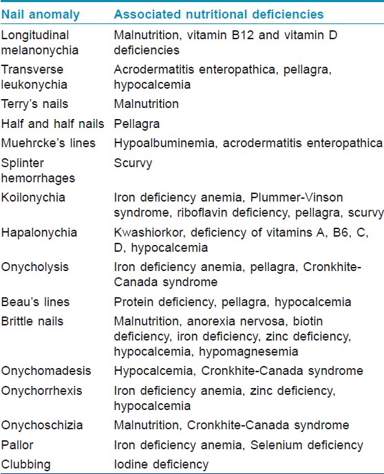
Table 1: Nail abnormalities and associated nutritional deficiencies
Nutritional supplements and the nails
A multitude of regimens exist for treatment of brittle nails, including biotin, application of essential fat acids, and ingestion of vitamin C, pyridoxine, fe, vitamin D, calcium, amino acids, and gelatin. [2] Conflicting evidence exists for the use of vitamin Due east to care for xanthous nail syndrome. [4] Other substances known to amend nail health merely defective objective testify include gelatin, cystine, L-methionine, keratin, collagen, pantothenic acid, salt, millet, yeast, chromium and rhodanates. [4]
Extended iron supplementation appears to subtract the brittleness of already brittle nails even in patients without overt iron deficiency. Studies have as well suggested that silicon supplementation can ameliorate the appearance of brittle nails. However, the best documented so far is the use of biotin to treat BNS. Clinical trial evidence exists and the beneficial effects unremarkably start after 2 to 3 months of supplementation. The optimal duration of the treatment has non been determined. [iv] Whether supplementation is legitimately correcting an underlying biotin deficiency or improvement in blast brittleness is through some other mechanism is yet to be elucidated. [2]
An exhaustive review on the role of vitamins and minerals in nail health ended that no evidence supports the use of vitamin supplementation for improving the nail health of well-nourished patients; breakable smash syndrome can do good from supplementation with high dose biotin or silicon; and lastly, adequate intake of vitamins and minerals facilitates smash wellness. [4]
References
| 1. | Tosti A, Piraccini BM. Biology of nails and nail disorders. In: Wolff K, Goldsmith LA, Katz SI, Gilchrest B, Paller AS, Leffell DJ, editors. Fitzpatrick's Dermatology In General Medicine. McGraw-Hill Companies Inc.; 2007. p. 778-94. [Google Scholar] |
| two. | Cashman MW, Sloan SB. Nutrition and nail illness. Clin Dermatol 2010;28:420-5. [Google Scholar] |
| 3. | Leonard PJ, Morris WP, Dark-brown R. Sodium, potassium, calcium and magnesium contents in nails of children with kwashiorkor. Biochem J 1968;110:22-3. [Google Scholar] |
| iv. | Scheinfeld Northward, Dahdah MJ, Scher R. Vitamins and minerals: Their function in nail health and affliction. J Drugs Dermatol 2007;6:782-7. [Google Scholar] |
| 5. | Solomons NW. On the assessment of zinc and copper nutriture in human being. Am J Clin Nutr 1979;32:856-71. [Google Scholar] |
| 6. | Longnecker MP, Stram Practise, Taylor PR, Levander OA, Howe M, Veillon C, et al. Apply of selenium concentration in whole blood, serum, toenails, or urine equally a surrogate mensurate of selenium intake. Epidemiology 1996;7:384-90. [Google Scholar] |
| 7. | Satia JA, King IB, Morris JS, Stratton Grand, White Due east. Toenail and plasma levels as biomarkers of selenium exposure. Ann Epidemiol 2006;16:53-8. [Google Scholar] |
| 8. | Sarkany R, Breathnach S, Morris A, Weismann G, Flynn P. Metabolic and Nutritional Disorders. In: Burns T, Breatnach S, Cox N, Griffiths C, editors. Rook'due south Textbook of Dermatology. John Wiley and Sons; 2010. p. 59.58-59.76. [Google Scholar] |
| 9. | Ghosn SH, Kibbi AG. Cutaneous manifestations of liver diseases. Clin Dermatol 2008;26:274-82. [Google Scholar] |
| 10. | Fawcett RS, Linford South, Stulberg DL. Smash abnormalities: Clues to systemic affliction. Am Fam Md 2004;69:1417-24. [Google Scholar] |
| 11. | Iorizzo M, Pazzaglia Thou, Piraccini B, Tullo S, Tosti A. Brittle nails. J Cosmet Dermatol 2004;three:138-44. [Google Scholar] |
| 12. | Gupta MA, Gupta AK, Haberman HF. Dermatologic signs in anorexia nervosa and bulimia nervosa. Arch Dermatol 1987;123:1386-ninety. [Google Scholar] |
| 13. | Cox N, Coulson I. Systemic disease and the skin. In: Burns T, Breatnach S, Cox Northward, Griffiths C, editors. Rook's Textbook of Dermatology. John Wiley and Sons; 2010. p. 62.57-62.58. [Google Scholar] |
| 14. | De Berker D, Baran R. Disorders of Nails. In: Burns T, Breatnach S, Cox N, Griffiths C, editors. Rook'due south Textbook of Dermatology. John Wiley and Sons; 2010. p. 65.1-65.17. [Google Scholar] |
| fifteen. | Marks J, Shuster Southward. Anaemia and peel disease. Postgrad Med J 1970;46:659-63. [Google Scholar] |
| 16. | Foti C, Cassano N, Palmieri VO, Portincasa P, Conserva A, Lamuraglia Chiliad, et al. Transverse leukonychia in astringent hypocalcemia. Eur J Dermatol 2004;14:67-8. [Google Scholar] |
| 17. | Simpson JA. Dermatological changes in hypocalcaemia. Br J Dermatol 1954;66:1-fifteen. [Google Scholar] |
| 18. | Reich H, Opitz One thousand, Bertram HP, Fegeler K. Successful zinc treatment of a severe instance of acrodermatitis enteropathica. Dtsch Med Wochenschr 1976;101:1724-6. [Google Scholar] |
| nineteen. | Kien CL, Ganther HE. Manifestations of chronic selenium deficiency in a child receiving total parenteral nutrition. Am J Clin Nutr 1983;37:319-28. [Google Scholar] |
| 20. | Ishida T, Himeno K, Torigoe Y, Inoue G, Wakisaka O, Tabuki T, et al. Selenium deficiency in a patient with Crohn'southward disease receiving long-term full parenteral nutrition. Intern Med 2003;42:154-7. [Google Scholar] |
| 21. | Al Hawsawi G, Pope E. Yellow nail syndrome. Pediatr Dermatol 2010;27:675-6. [Google Scholar] |
| 22. | Donald GF, Hunter GA, Gillam BD. Transverse leukonychia due to pellagra. Arch Dermatol 1962;85:530-one. [Google Scholar] |
| 23. | Cakmak SK, Gönül Chiliad, Aslan E, Gül U, Kiliç A, Heper AO. One-half-and-half nail in a case of pellagra. Eur J Dermatol 2006;sixteen:695-vi. [Google Scholar] |
| 24. | Ridley CM. Pigmentation of fingertips and nails in vitamin B12 deficiency. Br J Dermatol 1977;97:105-6. [Google Scholar] |
| 25. | Noppakun N, Swasdikul D. Reversible hyperpigmentation of peel and nails with white hair due to vitamin B12 deficiency. Arch Dermatol 1986;122:896-nine. [Google Scholar] |
| 26. | Niiyama S, Mukai H. Reversible cutaneous hyperpigmentation and nails with white hair due to vitamin B12 deficiency. Eur J Dermatol 2007;17:551-ii. [Google Scholar] |
| 27. | Carmel R. Pilus and fingernail changes in acquired and congenital pernicious anemia. Curvation Intern Med 1985;145:484-5. [Google Scholar] |

What Vitamin Deficiency Effects Nails,
Source: https://ijdvl.com/nails-in-nutritional-deficiencies/
Posted by: cooperabelity.blogspot.com
0 Response to "What Vitamin Deficiency Effects Nails"
Post a Comment